European Cetacean Bycatch banner loading

EUROPEAN CETACEAN BYCATCH CAMPAIGN
"Man is but a strand in the complex web of life"

Internal links buttons

HOME - SITE MAP - NEWS - CURRENT ISSUES - PHOTOS - ARCHIVE - CONTACT - LINKS - SEARCH

logomast7a.jpg


Gas bubble lesions in stranded cetaceans

Was sonar responsible for a spate of whale deaths after an Atlantic military exercise?

Nature – Vol 425 p575-6 9th October 2003

There are spatial and temporal links between some mass strandings of cetaceans — predominantly beaked whales — and the deployment of military sonar
1𔃁. Here we present evidence of acute and chronic tissue damage in stranded cetaceans that results from the formation in vivo of gas bubbles, challenging the view that these mammals do not suffer decompression sickness. The incidence of such cases during a naval sonar exercise indicates that acoustic factors could be important in the aetiology of bubble-related disease and may call for further environmental regulation of such activity.

Fourteen beaked whales were stranded in the Canary Islands close to the site of an international naval exercise (Neo Tapon, 2002) held on 24 September 2002. Strandings began about 4 hours after the onset of mid-frequency sonar activity. We necropsied eight Cuvier’s beaked whales (
Ziphius cavirostris), a Blainville’s beaked whale (Mesoplodon densirostris) and a Gervais’ beaked whale (Mesoplodon europaeus), six of which were very fresh. These animals showed severe, diffuse vascular congestion and marked, disseminated microvascular haemorrhages associated with widespread fat emboli within vital organs. Intravascular bubbles were present in several organs, although definitive evidence of gas embolism in vivo is difficult to determine after death 4. No pathogenic bacteria were isolated from the carcasses.

These lesions are consistent with acute trauma due to
in vivo bubble formation resulting from rapid decompression (as occurs in decompression sickness)4,5. Bubble formation in response to sonar exposure might result from behavioural changes to normal dive profiles (such as accelerated ascent rate), causing excessive nitrogen supersaturation in the tissues (as occurs in decompression sickness); alternatively, bubble formation might result from a physical effect of sonar on in vivo bubble precursors (gas nuclei) in nitrogen-supersaturated tissues6,7.

The beaked whales found in the Canary Islands are not the only stranded cetaceans to provide evidence of bubble-associated tissue injury. In strandings that occurred in Britain between October 1992 and January 2003, three out of 24 Risso’s dolphins (
Grampus griseus), three out of 342 common dolphins (Delphinus delphis) and one out of 1,035 harbour porpoises (Phocoena phocoena) necropsied, as well as the only Blainville’s beaked whale studied, contained gas bubbles in their blood vessels and gas-filled cavities in their parenchymous organs.

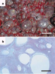
The livers of these animals were the most consistently affected organ, with macroscopic gas-filled cavities (diameter, 0.2𔃄.0 cm) occupying 5󈟆% of the volume (
Fig. 1) and having variable degrees of fibrotic encapsulation. Intrahepatic spherical non-staining cavities (gas bubbles) of diameter 50� mm were associated with compression of hepatic tissue, distension of portal blood vessels, and sometimes with haemorrhage, acute hepatocellular necrosis or fibrosis, indicating that this damage was inflicted before death. One of the D. delphis specimens also had bilateral acute renal infarcts associated with gas bubbles.

The cavitary lesions described here are new to marine-mammal pathology. Their presence in fresh carcasses in the absence of bacterial isolates, and the apparent progression through the stages of pericavitary fibrosis, are inconsistent with decompositional bubbles from bacterial activity. The coexistence of ante mortem gas bubbles and gasfilled fibrosed cavities suggests that
in vivo bubble formation and expansion is the proximate aetiology of this disease process. Bubble formation may either have initiated in the liver and kidneys (‘autochthonous’ bubble formation) or in other tissues (fatty tissue, for example) before haematogenous transfer to the liver and kidneys as gas emboli.

Figure 1 Gas-filled cavities in the liver of a stranded common dolphin

(Delphinus delphis).

a, Cut surface of the liver,
showing that cavitary lesions have
extensively replaced the normal tissue.


Scale bar, 10mm.


b, Photomicrograph of liver section,
showing multiple cavities
(gas bubbles) within the portal tracts
and hepatic parenchyma.

Scale bar, 750mm.


Nitrogen bubbles and emboli can develop in decompression sickness in humans and experimental animals as a result of expansion of pre-existing gas nuclei within nitrogen-supersaturated tissues
5. Anatomical, physiological and behavioural adaptations may mitigate against in vivo formation of nitrogen bubbles in marine mammals8󈝸, although there is empirical evidence of nitrogen supersaturation in cetaceans8.
Some deep-diving species are predicted to undergo up to 300% nitrogen tissue supersaturation
7.
Static diffusion in nitrogen-supersaturated tissues is therefore a plausible mechanism for bubble development and is consistent with a greater prevalence of cases in deep-diving species such as Risso’s dolphins and beaked whales.

Further investigation is needed into the physical and behavioural effects on cetaceans exposed to sonar, and the relation of these effects to bubble growth
in vivo and to strandings. Necropsies should aim to compare fat and gas emboli in stranded cetaceans suspected of having been exposed to sonar with results from unexposed stranded controls. In a wider conservation sense, our findings need to be taken into account in considering the regulation and limitation of the adverse impact of anthropogenic sonar on cetaceans.

P.D. Jepson*, M.Arbelo†, R. Deaville*, I. A. P. Patterson‡, P. Castro†, J. R. Baker§, E. Degollada†, H.M. Ross‡, P.Herráez†, A. M. Pocknell*, F. Rodríguez†, F. E.Howie||, A. Espinosa†, R. J. Reid‡, J. R. Jaber†, V. Martin†, A. A. Cunningham*, A. Fernández†

*
Institute of Zoology, Zoological Society of London,
Regent’s Park, London NW1 4RY, UK
Histology and Pathology Unit, Institute for
Animal Health, Veterinary School, Montana
Cardones-Arucas, University of Las Palmas de
Gran Canaria, Gran Canaria, Spain
e-mail: afernandez@dmor.ulpgc.es
Wildlife Unit, SAC Veterinary Science Division
(Inverness), Drummondhill, Stratherrick Road,
Inverness, IV2 4JZ, UK
§ Department of Veterinary Pathology, University of
Liverpool, Leahurst, Neston,Wirral CH64 7TE, UK
|| SAC Veterinary Science Division (Edinburgh),
Allan Watt Building, Bush Estate, Penicuik,

Edinburgh EH26 0QE, UK


Referemces
1. Simmonds, M. P. & Lopez-Jurado, L. F. Nature 337, 448 (1991).
2. Frantzis, A.
Nature 392, 29 (1998).
3. US Dept Commerce & US Navy
Joint Interim Report: Bahamas

Marine Mammal Stranding Event of 15󈝼 March 2000

http://www.nmfs.noaa.gov/prot_res/overview/
Interim_Bahamas_Report.pdf (2001).
4. Knight, B.
Forensic Pathology 2nd edn (Arnold, London, 1996).
5. Francis, T. J. R. & Mitchell, S. J. in
Bennett and Elliott’s
Physiology and Medicine of Diving
(eds Brubakk, A. O. &
Neuman, T. S.) 530� (Saunders, Philadelphia, 2003).
6. Crum, L. & Mao, Y.
J. Acoust. Soc. Am. 99, 2898� (1996).
7. Houser, D. S., Howard, R. & Ridgway, S. H.
J. Theor. Biol. 213,
183� (2001).
8. Ridgway, S. H. & Howard, R.
Science 206, 1182� (1979).
9. Ponganis, P. J., Kooyman, G. L. & Ridgway, S. H. in Bennett and
Elliott’s Physiology and Medicine of Diving
(eds Brubakk, A. O. &
Neuman, T. S.) 211� (Saunders, Philadelphia, 2003).
10. Harrison, R. J. & Tomlinson, D.W.
Proc. Zool. Soc. Lond. 126,
205� (1956).
11. Ridgway, S. H. & Howard, R.
Science 216, 651 (1982).
12. Falke, K. J.
et al. Science 229, 556� (1985).


Top

gasfilledcavititesinstrandedcetaceansandphotomicrograph.jpg